


Sir, Lipodystrophy can result in disfigurement in view of the progressive loss of fat. Nodular swellings of panniculitis often precede the onset of overt lipodystrophy. The patient reported here has had a documented panniculitis in the past, with positive autoantibodies.
Mrs A was a 37‐yr‐old lady who presented for evaluation of her facial disfigurement. She complained of loss of superficial tissue resulting in furrowing of the face and abnormal contours of her limbs. Her history dates back to 1987 when she noticed multiple nodular swellings predominantly over the back, shoulders, abdomen and occasionally over the extremities and face. This was biopsied at that time and found to be consistent with a panniculitis, septal in nature. These recurred in crops over the same region and disappeared within a few months. Between 1992 and 1994 she noticed progressive loss of superficial tissue over the face and extremities. There was patchy loss of tissue over the trunk. Subsequent to 1994 there was no progression. There was no history of joint pains, oral ulcers or a malar rash. She denied having had drying of the eyes or mouth or having had painful muscles or motor weakness at any time. There was no family history of a similar illness. She was not a known diabetic or hypertensive on presentation.
Clinical examination revealed a middle‐aged lady who was comfortable at rest. Her vital signs were normal. Her face was hollowed to the contours of her facial bones. Muscle contours over her extremities were clearly demarcated. Her calves demonstrated pseudohypertrophy due to loss of fat (Fig. 1). Besides the bulk of the muscle, only the tendon remained. Over the trunk, however, there was patchy loss of subcutaneous tissue. The upper limbs were relatively spared. General physical examination was normal, and there was no significant lymph node enlargement. Examination of the eyes and teeth showed these to be normal. Currently there are no nodular eruptions on examination. Systemic examination is normal except for a soft hepatomegaly, palpable 2 cm below the costal margin.
Mrs A's complete blood counts were normal. Her erythrocyte sedimentation rate was 30 mm at 1 h. Her liver function tests and serum creatinine were normal. Her lipid profile revealed triglycerides of 4.91 mmol/l. Her low‐ and high‐density lipoprotein cholesterol and total cholesterol were normal. Her fasting plasma glucose was 9.76 mmol/l, post‐prandial plasma glucose was 13.7 mmol/l, confirming her diabetic status. Her routine urine microscopy was normal. Her thyroid function tests were normal. The serum albumin:globulin ratio and their levels were normal. There was no proteinuria detected. Her liver function tests were normal. Her serum complement was normal and her lupus erythematosus cell preparation on peripheral blood was negative. The antinuclear antibody was weak positive and antibody levels to double‐stranded DNA were elevated (41.8 U/ml, normal<30 U/ml). Her skin biopsy revealed no specific lesion.
Mrs A presented with the clinical syndrome of lipodystrophy associated with an antecedent panniculitis. Morphea was considered, but excluded in view of the negative skin biopsy.
The following associations have been described between connective tissue disease and panniculitis. Lupus panniculitis, scleroderma panniculitis, dermatomyositis panniculitis, connective tissue panniculitis and localized lipoatrophy [1]. Sjögren's syndrome and isolated low complement levels have been described in patients with lipodystrophy [2]. Mrs A has not had clinical features of the above autoimmune diseases. Her serum complement was normal.
Lupus panniculitis occurs in 2–3% of patients with systemic lupus erythematosus [3, 4]. Handsfield et al. [5] describe an Indian woman who had a lipodystrophy following a panniculitis and was antinuclear antibody positive. She did not have antibodies to double‐stranded DNA. The panniculitis preceding a lipoatrophy seems to suggest that the lipoatrophy is a focal expression of connective tissue disease [6].
Lipodystrophy has been associated with diabetes mellitus [6–8]. Congenital total lipodystrophy is an autosomal recessive disorder and presents as a clinical syndrome with complete absence of adipose tissue, nephropathy, mental retardation, precocious puberty, acanthosis nigricans, insulin resistance, diabetes mellitus and hypertriglyceridaemia [7]. Insulin resistance may occur in the acquired variants of lipodystrophy as well [7]. Insulin‐dependent diabetes mellitus has been described preceding the onset of lipodystrophy [6, 9].
Mrs A has the features of a late‐onset acquired lipodystrophy with mild diabetes mellitus associated with hypertriglyceridaemia with no features of end organ damage secondary to diabetes mellitus. She is on a diet for her diabetes. The mild diabetes of recent onset is unlikely to be the cause of the lipodystrophy in this case.
The case describes an acquired lipodystrophy with accompanying diabetes mellitus and positive autoantibodies. This association without clinical features of connective tissue disease suggests that lipodystrophy is part of a clinical spectrum, rather than a distinct clinical entity.
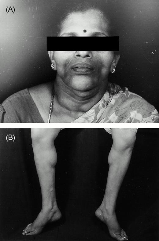
(A) The face of a patient with lipodystrophy reveals the loss of adipose tissue and bony and muscular contours are seen. (B) The patient's calves demonstrate the muscular contours demarcated due to the loss of fat.
Correspondence to: A. M. Cherian.
References
Donaton AS, Francisco RN. Association of partial lipodystrophy and Sjögren's syndrome.
Diaz JE, Horatius RJ, Alarcon SD et al. Systemic lupus erythematosus, presenting as panniculitis (lupus profundus).
Tuffanelli DL. Lupus erythematosus panniculitis (Profundus). Clinical and immunologic studies.
Handsfield SEJ, Stephens CJM, Mayou BJ, Black MM. The clinical spectrum of lipoatrophic panniculitis includes that of connective tissue panniculitis.
Kohli V, Shankar RR, Singh M. Lipodystrophy in association with unstable diabetes mellitus.
Julie KB, Sandy SM, Aditya KG, John TH, James ER. Lipoatrophic panniculitis: a possible autoimmune inflammatory disease of fat.

Comments